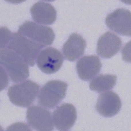
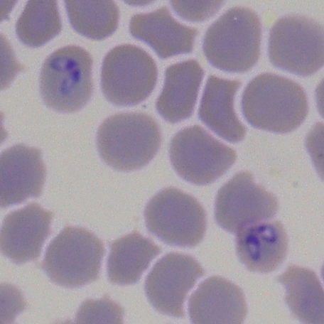
Theileria equi and Babesia caballi in vitro culture
Equine piroplasmosis (EP) is an infectious disease of equids caused by the intraerythrocytic protozoan parasites Babesia caballi (B. caballi) and Theileria equi (T. equi). The importance of this disease is the fact that, after the infection, horses can remain as chronic carriers of the parasites for a long time, being seropositive but showing clinical signs only in cases of immunosuppression or intense exercise. EP involves serious economic losses for the equine industry due to the restrictions in the export of horses to countries free from EP.
For the diagnosis of EP, both direct methods (visualization of parasites in blood smears, PCR and in vitro culture) and indirect or serological methods (cELISA, complement fixation test and indirect immunofluorescence assay) can be used.
The in vitro culture of these parasites was described in 1987. The first data on the specific culture of B. caballi and T. equi were published in 1993 and 1994, respectively. Since then and until 2002, several studies have been published in order to achieve an improvement of this technique and to identify carrier horses.
Recently, this technique has been implemented in our laboratory thanks to a collaboration with the USDA-APHIS diagnostic laboratory (Iowa, EEUU), achieving to culture isolates from field strains of both parasites. Currently, this technique is performed in very few laboratories, since very specific conditions are needed for the growth of parasites.
In order to carry out T. equi and B. caballi in vitro culture, it is necessary to have a clean room designed exclusively for that purpose. Blood of suspect horses used for the initiation of cultures should be collected in tubes with EDTA as an anticoagulant. In the laboratory, the erythrocytes are washed several times through centrifugation and subsequent resuspension of the pellet with sterile PBS. Blood plasma and white cells must be removed after each wash.
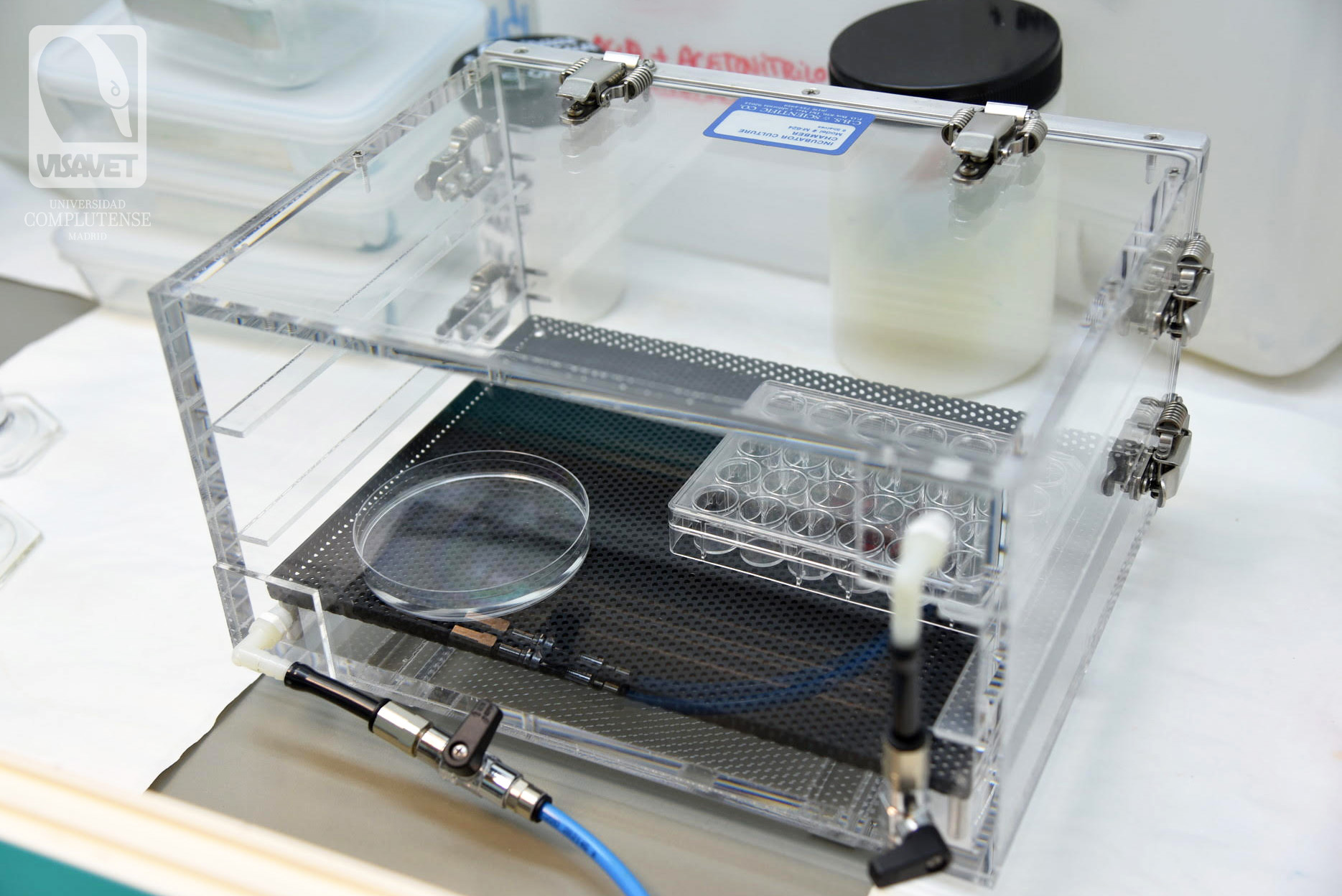
Washed erythrocytes are incubated in different cell culture plates with two specific mediums, one for T. equi and one for B. caballi. The specific medium is done in a class II biosafety cabinet and among other components, aminoacids, enzymes, antibiotic/antimycotics and a pH stabilizing buffer are used. Both plates are placed in different modular incubator chambers, which must be filled with a gas mixture composed of different concentrations of O2, CO2 and N (what is termed “blood gas mixture”). This gas mixture is allowed to flow for 1 min to get a full filled chamber.
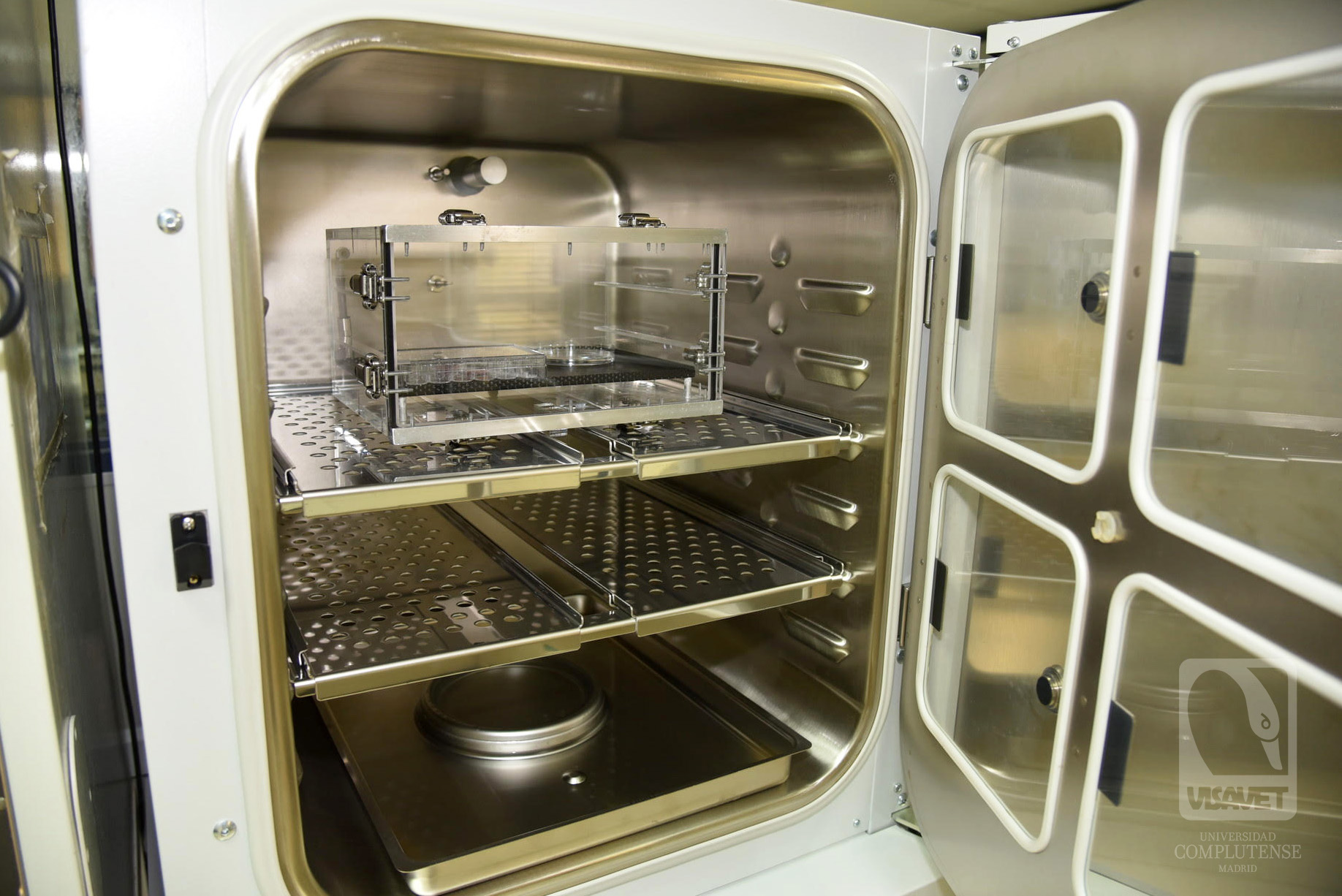
Finally, both modular incubator chambers are kept within a humidified CO2 incubator in order to get a controlled environment. Once the cultures have been initialized, the specific medium should be changed daily and the growth and multiplication of the parasites should be monitored by visualizing them in a blood smear at least once a week. The growth time of the parasites will depend on the initial existing parasitemia percentage. This growth time can vary from 2 weeks to one month.
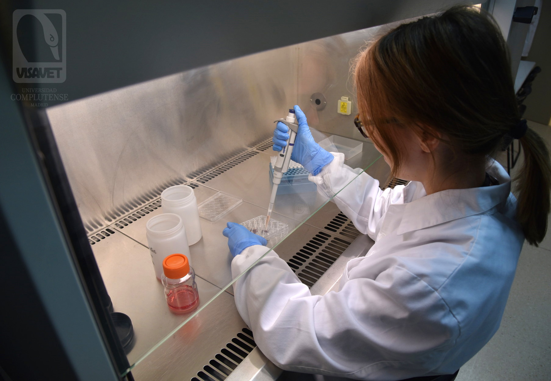
Although the main goal to be achieved with the in vitro culture is its use as an additional method for the identification of EP carrier horses, it is also a technique for other purposes such as the evaluation of the in vitro effect of antiparasitic drugs, isolation, sequencing and molecular characterization of field strains and the production of specific antigens for the development of new diagnostic methods and vaccines.
How to send us samples for informative analyses
The Equine Health Surveillance Unit (SEVISEQ) from the VISAVET centre is a Laboratory authorised for the diagnosis of Equine Piroplasmosis by cELISA, IFAT, CFT and PCR samples (prior to exportation or movements and from horses with a suspicion of Equine Piroplasmosis and/or with clinical signs).
In order for us to carry out these tests, serum samples must be identified with the transponder number or UELN number as well as the number of the premises where the horse resides.
For further information and the consultation of questions regarding Equine Piroplasmosis please contact us:
VISAVET Health Surveillance Centre
Complutense University Madrid (Spain)
Tel.: (+34) 913944096
seviseq@ucm.es
Bibliography
- Anon. 2014. Equine piroplasmosis. OIE Manual of Diagnostic Tests and Vaccines for Terrestrial Animals. Chapter 2.5.8.
- Zweygarth E, Vanniekerk C, Just MC, Dewaal DT. 1995. In-Vitro Cultivation of a Babesia Sp from Cattle in South-Africa. Onderstepoort J Vet. 62(2):139-42.
- Holman PJ, Frerichs WM, Chieves L, Wagner GG.. 1993. Culture confirmation of the carrier status of Babesia caballi-infected horses. J Clin Microbiol. 31(3):698-701.
- Posnett ES, Ambrosio RE.. 2009. DNA probes for the detection of Babesia caballi. Parasitology. 102(3):357-65.
- Zweygarth E, van Niekerk CJ, de Waal DT. 1999. Continuous in vitro cultivation of Babesia caballi in serum-free medium. Parasitol Res. 85(5):413-6.
- Zweygarth E, Lopez-Rebollar LM, Nurton J, Guthrie AJ. 2002. Culture, isolation and propagation of Babesia caballi from naturally infected horses. Parasitol Res. 88(5):460-2.
- Schein E, Rehbein G, Voigt WP, Zweygarth E. 1981. Babesia equi (Laveran 1901) 1. Development in horses and in lymphocyte culture. Tropenmed Parasitol. 32(4):223-7.
- Zweygarth E, Just MC, DeWaal DT. 1997. In vitro cultivation of Babesia equi: Detection of carrier animals and isolation of parasites. Onderstepoort J Vet. 64(1):51-6.
- Holman PJ, Chieves L, Frerichs WM, Olson D, Wagner GG. 1994. Babesia equi erythrocytic stage continuously cultured in an enriched medium. J Parasitol. 80(2):232-6.
- Zweygarth E, Just MC, van Niekerk C, de Waal DT. 1997. In vitro cultivation of Babesia equi and its applications. Trop Anim Health Pro. 29(4):60s-s.
- Holman PJ, Hietala SK, Kayashima LR, Olson D, Waghela SD, Wagner GG. 1997. Case report: field-acquired subclinical Babesia equi infection confirmed by in vitro culture. J Clin Microbiol. 35(2):474-6.
- Alhassan A, Govind Y, Tam NT, Thekisoe OM, Yokoyama N, Inoue N, et al. 2007. Comparative evaluation of the sensitivity of LAMP, PCR and in vitro culture methods for the diagnosis of equine piroplasmosis. Parasitol Res. 100(5):1165-8.


