

Strongylosis
Etiology
Intestinal nematodes belonging to the family Strongylidae, which is divided into two groups: the subfamily Strongylinae (“large strongyles”) and the subfamily Cyathostominae (“small strongyles”). Within the subfamily Strongylinae stands out the genus Strongylus (S. vulgaris, S. edentatus and S. equinus).
Epidemiology
The nematodes of the family Strongylidae present a direct biological cycle with two phases: exogenous and endogenous. Exogenous phase begins with the non embryonated eggs of adult strongyles in the feces of horses. In the environment L1 is formed and hatched, which molts to L2 and after to L3 in about a week. The infective stage is L3, which are sorrounded by a durable sheath that protects it from low temperaturas but does not allow them to feed. The L3 leaves the manure and goes to the grass. Horses become infected by ingesting the L3 in pasture.
The endogenous phase starts inside the host, where the L3 can migrate to organs or not. S. vulgaris L3 reaches the large intestine and molts to L4, which ascends to the anterior mesenteric artery and molts to L5 or preadult. S. equinus L3 reaches the large intestine and forms nodules, inside which it molts to L4. L4 goes to the liver and after to the pancreas, where it molts to L5. S. edentatus L3 reaches the large intestine and then goes to the liver. In the liver, it forms liver nodules and molts to L4. Later, it migrates to the parietal peritoneum and molts to L5. In the three previous species, L5 returns to the large intestine. Adults are developed and females lay eggs that will come out in the faeces of the host.
Regarding the subfamily Cyathostominae, L3 invade the mucosa of the large intestine and form cysts, inside of which the L3 molt to L4. Finally, L4 larvae will become adults. L3 hypobiosis can occur: they stop their biological cycle, staying encysted in the mucosa to avoid unfavorable environmental conditions (winter in northern climates or summer in southern climates).
Pathogeny
Both adult and larval stages can cause damage to the host. The fixation of adult worms in the mucosa of the cecum or ventral colon results in a rupture of local vessels causing focal haemorrhages and even deep ulcers. Due to the migrating larvae of S. vulgaris can occur lesions such as thrombi, arteritis and hypertrophy of the anterior mesenteric artery medial layer. This is described as verminous arteritis and is associated with an increased incidence of colic. Migrating larvae of S. edentatus and S. equinus is related to a haemorrhagic and inflammatory lesion in the liver, pancreas and retroperitoneal tissues. At the end of the migratory period, all the Strongylus larvae return to the large intestine and form nodules within the gut wall. These nodules often are filled with purulent material. Rupture of the cysts during larval emergence of subfamily Cyathostominae results in lesions of focal oedema, congestion, and haemorrhage, with accompanying leakage of tissue fluids.
Clinical signs
Strongylosis is characterized by nonspecific signs such as weight loss and poor growth, rough hair coat, and compromised performance. Experimental infections caused pyrexia, tachycardia, anorexia, and diarrhoea. Larval S. vulgaris infections are associated with colic. In the case of subfamily Cyathostominae, larval cyathostominosis syndrome can occur. This syndrome is caused by emergence or large numbers of encysted cyathostomins and it is characterized by diarrhoea, rapid weight loss and marked hypoproteinemia.
Diagnosis
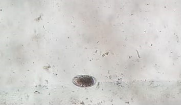
The most common diagnostic technique is coprologic examination for the visualization of the adult parasites eggs. Cyathostomins eggs cannot be differentiated from large strongyle eggs. In horses with strongylosis we can find hematological (anemia, leukocytosis and eosinophilia) and biochemical (hypoalbuminemia) findings. On the other hand, both serological techniques (ELISA) and molecular biology techniques (PCR and RLB) have been described for the detection of several species of small and large strongyles. Recently, a real-time PCR assay has been developed for the detection and semiquantification of S. vulgaris DNA in faecal samples.
Treatment
In general, benzimidazoles, macrocyclic lactones (ivermectin and moxidectin) and tetrahydro-pyrimidines (pyrantel) are effective against adult strongyles. Only fenbendazole and moxidectin would be effective to encysted larvae of small strongyles. In addition to these two anthelmintics, ivermectin could be used against migrating larvae of large strongyles.
Prevention and control
Prevention and control: The daily administration of pyrantel can be effective to the control of large strongyles. However, resistance to pyrantel has been demonstrated in the case of cyathostomins. The best form of control is deworming of horses twice a year, at intervals of 6 months. Duddingtonia flagrans fungi, which grows in the sheep, cattle and horses faeces, would be a biologic control since it is a natural predator and kill free-living larvae.
Public Health Considerations
These nematodes have no zoonotic potential and cannot infect any domestic animals other than equids.
References
- Equine Infectious Diseases. 2014 Elsevier Inc. ISBN: 978-1-4557-0891-8
- Studies on the epidemiology of Strongylus vulgaris of the horse.
- The Life Cycle, Pathogenisis and Epidemiology of S. vulgaris in the horse.
- Small strongyles: recent advances.
- Epidemiology and control of equine strongylosis in Newmarket.